-
Home
-
About JCTR
-
Gold Open Access
-
Issues
-
Editorial board
-
Author guidelines
-
Publication fees
-
Online first
-
Special issues
-
News
-
Publication ethics
-
Partners
-
Submit your manuscript
-
Submit your review report
-
Editorial Office
-

This work is licensed under a Creative Commons Attribution-NonCommercial 4.0 International License. ISSN print: 2382-6533 ISSN online: 2424-810X
Volume 5 Issue 2
Circulating plasma microRNA-126, -145 and -155 and their association with atherosclerotic plaque characteristics
Evija Knoka, Karlis Trusinskis, Mairita Mazule, Ieva Briede, William Crawford, Sanda Jegere, Indulis Kumsars, Inga Narbute, Dace Sondore, Aivars Lejnieks, Andrejs Erglis
Knoka et al., J Clin Transl Res 2019; 5(2): 2
Published on January 13, 2020
Abstract
Background and Aim: Circulating microRNAs (miRNAs) have been identified as biomarkers for several diseases. Dysregulation of miRNA-126, -145 and -155 have been shown to be associated with atherosclerotic lesion formation. The aim of this study was to evaluate the association between atherosclerosis-related miRNAs and unfavourable atherosclerotic plaque characteristics.
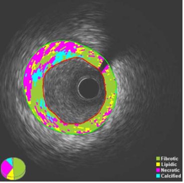
Methods and Results: Forty patients with stable coronary artery disease (CAD) admitted for elective percutaneous coronary intervention (PCI) were enrolled in a prospective study. After PCI, intravascular ultrasound (IVUS) and iMAP-IVUS analysis was performed to assess the proportion of fibrotic, necrotic, lipidic and calcific tissue within atherosclerotic plaques. Total RNA was isolated from plasma to evaluate the expression of circulating miRNA-126, miRNA-145 and miRNA-155. Plasma lipid and glucose metabolism-related variables were measured to determine any association with plaque characteristics or miRNA expression. Expression of miRNA-126 was negatively correlated with plaque fibrotic tissue (r=-0.28; p=0.044), while positively correlated with plaque necrotic tissue (r=0.31; p=0.029) and necrolipidic tissue (r=0.31; p=0.031). MiRNA-145 was positively correlated with plaque lipidic (r=0.32; p=0.023) and necrolipidic tissue (r=0.31; p=0.029). Patient age was associated with plaque fibrotic tissue (r=-0.41; p=0.005), necrotic tissue (r=0.33; p=0.022), and lipid content (r=0.33; p=0.022). HDL-C was positively correlated with plaque necrotic (r=0.28; p=0.042) and calcific (r=0.28; p=0.044) tissue volume. Calcific tissue volume was positively correlated with C-peptide (r=0.34; p=0.033). After multivariate logistic regression analysis, both miRNA-126 and miRNA-145 expression were associated with increased necrolipidic tissue content (β=0.34; p=0.050; and β=0.35; p=0.037 respectively).
Conclusions: Expression of miRNA-126 and miRNA-145 were associated with increased plaque necrolipidic tissue content.
Relevance for patients: Although further research is needed to support the study data, miRNA-126 and miRNA-145 may serve as potential plaque vulnerability biomarkers in the future.
DOI: http://dx.doi.org/10.18053/jctres.05.201902.002
Author affiliation
1 Latvian Centre of Cardiology, Pauls Stradins Clinical University Hospital, Latvia
2 Riga Stradins University, Latvia
3 University of Latvia, Latvia
4 Riga East Clinical University Hospital, Latvia
5 University of Oxford, United Kingdom
*Corresponding author
Evija Knoka, Pauls Stradins Clinical University Hospital, Pilsonu Street 13, Riga, LV-1002, Latvia.
Tel: +371 29376907
E-mail: evija.knoka@gmail.com
Handling editor:
Michal Heger
Department of Pharmaceutics, Utrecht University, the Netherlands
Department of Pharmaceutics, Jiaxing University Medical College, Zhejiang, China