-
Home
-
About JCTR
-
Gold Open Access
-
Issues
-
Editorial board
-
Author guidelines
-
Publication fees
-
Online first
-
Special issues
-
News
-
Publication ethics
-
Partners
-
Submit your manuscript
-
Submit your review report
-
Editorial Office
-

This work is licensed under a Creative Commons Attribution-NonCommercial 4.0 International License. ISSN print: 2382-6533 ISSN online: 2424-810X
Volume 5 Issue 3
Investigation of quantitative susceptibility mapping (QSM) in diagnosis of tuberous sclerosis complex (TSC) and assessment of associated brain injuries at 1.5 Tesla
Lei Zhang, Hongqiang Xue, Tao Chen, Hongzhe Tian, Xiaohu Wang, Xiaocheng Wei, Huawen Zhang, Hui Ma, Zhuanqin Ren
Zhang et la., J Clin Transl Res 2020; 5(3): 3
Published online: March 11, 2020
Abstract
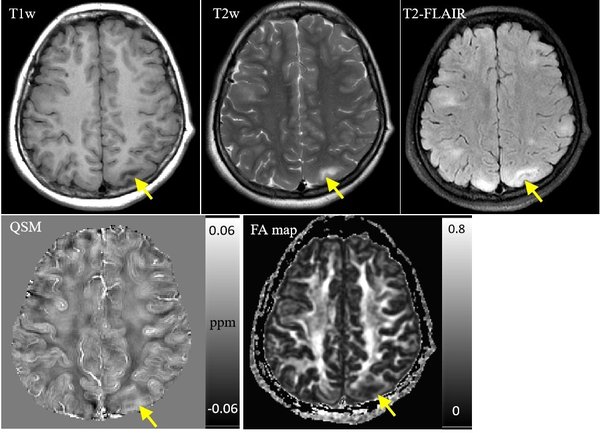
Background and Aim: Tuberous sclerosis complex (TSC) is a rare disease with serious clinical consequences, such as mental deficiency and epilepsy. The pathological changes of TSC include demyelination and subependymal calcified nodules. Quantitative susceptibility mapping (QSM) is a newly developed imaging technique which is capable of quantitatively measuring the susceptibility induced by iron deposition, calcification and demyelination. The aim of this study was to investigate the use of QSM in detecting the subependymal nodules and assessing brain tissue injuries induced by cortical/subcortical tubers in TSC patients.
Methods: Twelve clinically confirmed TSC patients and fifteen gender and age matched healthy subjects underwent measurement with conventional magnetic resonance imaging (MRI) sequences, diffusion tensor imaging (DTI) and QSM. The TSC patients further underwent a computed tomography (CT) scan. Considering CT as the ground truth, the detection rates of subependymal nodules using conventional MR images and QSM was compared by the paired Chi square test, and the sensitivity, specificity were computed. The Bland-Altman test and independent t test was performed to compare the susceptibility of cortical/subcortical regions from QSM and fractional anisotropy (FA) values from DTI between the patient and control groups, pearson correlation was performed to examine the correlation between the susceptibility and FA values.
Results: QSM was better in detecting subependymal calcified nodules compared to conventional MR sequences (X2 = 40.18, p < 0.001), QSM achieved a significantly higher sensitivity of 98.3% and a lower specificity of 50%, which was compared with conventional MR sequences (46.7%, 75%, respectively). The susceptibility value of cortical/subcortical tubers in TSC patients was significantly higher than those in control group (t = 9.855, p < 0.001), while FA value was lower (t = -8.687, p < 0.001). Pearson correlation test revealed negative correlation between susceptibility and FA values in all participants (r = -0.65, p < 0.001).
Conclusions: QSM had similar ability in TSC compared to CT and DTI. QSM may provide valuable complementary information to conventional MRI imaging, and may simplity imaging of patients with TSC.
Relevance for patients: This study shows the feasibility of QSM to detect subependymal calcified nodules. It may provide quantitative information of white matter damage of tuberous sclerosis patients.
DOI: http://dx.doi.org/10.18053/jctres.05.202003.003
Author affiliation
1 Department of Radiology, Baoji Hi-Tech People's Hospital, Baoji, Shaanxi, China
2 Department of Radiology, Baoji Center Hospital, Baoji, Shaanxi, China
3 GE Healthcare, Beijing, China
4 Department of Radiology, Nuclear Industry 215 Hospital of Shaanxi Province, Xianyang, Shaanxi, China
*Corresponding author
Zhuanqin Ren
Department of Radiology, Baoji Center Hospital, No. 8 Jiang Tan Road, Baoji 721008, Shaanxi, P.R. China.
E-mail: renzhuanqin@163.com
Tel: +86-13892451698.
Hui Ma
Department of Radiology, Baoji Hi-Tech People's Hospital, No. 19 He Xie Road, Baoji, 721013, Shaanxi, P.R. China.
E-mail: mahui198023@163.com
Tel: +86-13991575903
Handling editor:
Michal Heger
Department of Pharmaceutics, Utrecht University, the Netherlands
Department of Pharmaceutics, Jiaxing University Medical College, Zhejiang, China